- Feb 19, 2024
The Science Behind BAEPs – Brainstem Auditory Evoked Potentials! Part 2
- Cesia M. Alvarez & Faisal Jahangiri
- 0 comments
Cesia M. Alvarez & Faisal R. Jahangiri
In the initial installment of this blog series discussing BAEPs, auditory potentials are utilized by IONM experts to assess and interpret auditory signals and their corresponding cortical pathways. Each component, from the physical features of the outer ear to the complex subcortical pathways within the temporal lobe - responsible for auditory processing in the brain - plays a critical role in receiving, transcribing, and further cortical processing of these neuronal signals [2].
There are three types of auditory responses: Electrocochleogram (ECoch), Auditory Nerve Compound Action Potentials (AN-CAP), and Brainstem Auditory Evoked Potentials/Responses (BAEP/BAER) the latter being the focus of this discussion. Each response focuses on a specific auditory aspect of the signal or pathway processing location. ECoch, which has an amplitude of around 2-20 µV, can be subdivided into three categories: Cochlear Microphonic (CM), which stimulates the Cochlea by acoustic means; Summation Potential (SP), which involves the same mode of stimulation as CM but also has to be consistent with the presence of cochlear perfusion, and Compound Action Potential (CAP) which is the activation of the auditory nerve by the stimulation of myelinated segments of that nerve producing N1 which is identical to wave I of a BAEP (N1 being the event-related potential (ERP) that is usually considered for the evaluation of auditory processing [3,4]. AN-CAPs, on the other hand, are signals that are directly recorded from the auditory nerve, and the near-field N1 response has the same latency as wave II of a BAEP, but it’s not wave II [3]. Meanwhile, BAEPs and BAERs are signals recorded by three electrodes placed on the scalp and two other electrodes near the ear. These signals result in the identification of waves I, II, III, IV, V, VI, and VII (Fig 1.), as previously mentioned in the first blog (Fig. 1) [3].
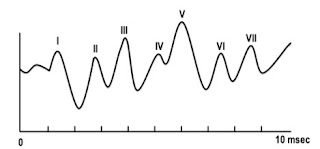
Figure 1. Shows the BAEP waves with their respective numbers along the auditory nerve pathway. Surgical Neurophysiology 2nd Ed [3].
To fully comprehend the significance of each peak, one must familiarize oneself with the neural map of the auditory nerve pathway, a component of the vestibulocochlear nerve (the eighth cranial nerve). The pathway is divided into two nerve branches: the vestibular nerve, responsible for balance, and the cochlear nerve, responsible for hearing [3]. The vestibular nerve branches from the eighth cranial nerve and supplies the semicircular canals in the inner ear. It then synapses with the vestibular nucleus located in the fourth ventricle of the pons and medulla [3]. On the other hand, the cochlear nerve originates from the cochlea and joins the vestibular nerve branch to form the vestibulocochlear or eighth cranial nerve [3]. As previously stated, each peak wave identified by a number represents a location along the auditory nerve pathway. A helpful diagram illustrating the localization and delineation of these peak wave generators can be found in Fig. 2.
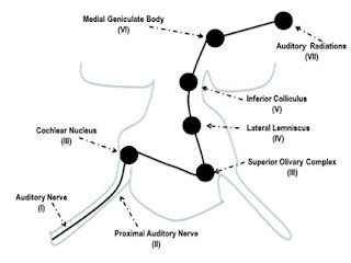
Figure 2. A diagram showing the localization of peaks I – VII along the auditory nerve pathway. Surgical Neurophysiology 2nd Ed [3].
According to the above diagram, the auditory signal travels through various areas before reaching the auditory cortex. These areas include the auditory nerve, cochlear nucleus, superior olivary complex, lateral lemniscus, inferior colliculus, and medial geniculate body. It is worth noting that all these cortical regions are crucial for processing and filtering the final auditory response [1,3].
Sources:
Irving, R., and J. M. Harrison. “The Superior Olivary Complex and Audition: A Comparative Study.” Journal of Comparative Neurology, JCN 130, no. 1 (May 1967): 77–86. https://doi.org/10.1002/cne.901300105.
Purves D, Augustine GJ, Fitzpatrick D, et al., editors. Neuroscience. 2nd edition. Sunderland (MA): Sinauer Associates; 2001. The Auditory Cortex. Available from: https://www.ncbi.nlm.nih.gov/books/NBK10900/#.
Surgical Neurophysiology – A reference guide to intraoperative neurophysiological monitoring (IONM), 2nd edition by Faisal R. Jahangiri, MD.
Wang, An Li, André Mouraux, Meng Liang, and Gian Domenico Iannetti. “The Enhancement of the N1 Wave Elicited by Sensory Stimuli Presented at Very Short Inter-Stimulus Intervals Is a General Feature across Sensory Systems.” PLOS ONE 3, no. 12 (December 12, 2008). https://doi.org/10.1371/journal.pone.0003929.