- Dec 27, 2024
Basis of Fear and its Effects: A Literature Review
- Sumana Karnala & Faisal Jahangiri
- 0 comments
Introduction
This literature review focuses on understanding the neural basis of fear and how it is processed, from the origin to the expression of fear behavior. Fear is a fundamental, archetypical emotion strongly associated with survival. From an evolutionary viewpoint, fear is a warning signal intended to establish a “fight or flight” response to extend an organism’s longevity. From a social standpoint, fear is often associated with anxiety and influences behavior with peers. Fear is omnipresent, whether regarding the smallest of a day’s concerns to Post Traumatic Stress Disorder–and therefore is necessary to be studied. The neural mechanisms underlying fear processing are intricate and just as fascinating, warranting a complex discussion.
Evolutionary Background and Foundations
Charles Darwin emphasized the phylogenetic continuity of emotions in that nonhuman primates and rodents have shown incredible similarity to human emotions, particularly fear and aggression (Darwin, 1872). This homogeneity indicates that fear had evolved in response to predation before conscious decision-making in threat-avoidant behavior. As a result of painful stimuli, animals have developed a need to be wary of threats to predict predator behavior subconsciously. Some emotions, like awe or guilt, may be unique to humans (Adolphs, 2013). These emotions require conscious analysis of alternate possibilities. For example, humans tend to feel awe when they encounter new experiences and guilt when they are aware of a better decision that could have been made. Animals cannot reflect upon this, although precursors to these emotions may be present (Adolphs, 2013). This interpretation establishes fear as a primal, archetypical emotion that can be evaluated through animal models, allowing for a better understanding of comparable fear in humans.
Classic theories for the definitions of fear have set a foundation for which fear pathways can be better analyzed. The James–Lange theory proposes that firstly, an emotional stimulus is encountered, inducing “peripheral physiological variations” that are then interpreted by the brain to be the emotion of fear (D’Hondt, 2010). For example, sensations such as increased heart rate because of seeing a fear-inducing stimulus would create the feeling of fear. In contrast, the Cannon-Bard theory suggests that encountering an emotional stimulus produces brain activity that simultaneously induces “bodily responses…and subjective feeling” (D’Hondt, 2010). Similarly, the Schachter-Singer theory states that fear is a union of physical arousal and interpretation based on the environment and circumstance (McLeod, 2023). For example, an emotional stimulus is encountered, followed by an increased heart rate and the conscious self-acknowledgment of fear and experiencing fear.
In addition to countless laboratory experiments and observations, technological advancements have allowed researchers to understand better the brain mechanisms inducing feelings of fear. For example, resting-state functional MRI has been utilized before and following fear conditioning to measure the variations between brain regions at rest and because of fear exposure (Greco, 2015). Positron emission tomography (PET) has also been used to measure levels of brain activity using a tracer, allowing for the tracking of metabolites associated with fear pathways (Bremner, 2011). With this newfound technology, scientists have been able to understand more than the psychology of fear and can now identify the changes in brain activity.
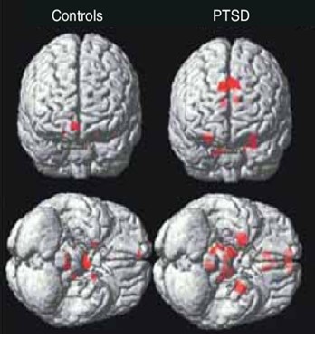
Figure 1. Functional MRI images. The fMRI image showcases the differences in brain region activity between a control patient exposed to neutral stimuli (left) and a PTSD patient reflecting on past traumatic memories (right). Areas such as the amygdala show heightened activation due to the processing of fear and the corresponding manifestation of physiological responses (Gordon, 2007).
Neural Circuitry of Fear
The amygdala, a crucial part of the limbic system, is a “passive output station for processing and plasticity” (Keifer Jr, 2015). This almond-shaped structure processes visual and auditory stimuli to determine what may be classified as dangerous (Amygdala, 2023). As a result of this neural plasticity, upon encountering a similar fear-inducing stimulus in the future, the amygdala can induce similar feelings, often interpreted as wariness. The hippocampus plays a role in memory, particularly in the “conditioning of fear to contextual information” (Kjelstrup, 2002). This association aids the prefrontal cortex in modulating fear by distinguishing between safe and harmful situations. The hypothalamus then allows for the physical manifestation of emotions of fear. By sending signals through the autonomic nervous system, the hypothalamus causes the pituitary glands to release hormones (Ackerman, 1992). As a result, fear results in the physiological sensations of fast heart rate, shallow breathing, and sweating.
Dysregulation of Fear Processing in Mental Disorders
Excessive fear is a significant component of anxiety disorders, often increasing “amygdala activation” because of “disorder-relevant stimuli,” such as PTSD and phobias (Shin, 2009). This altered fear circuitry contributes to heightened feelings of worry and the manifestation of harmful physiological responses, such as heart palpitations. The association of traumatic events with fear and wariness causes neural changes associated with experiencing symptoms, even in the absence of the same traumatic stimuli (Shin, 2009). In other words, a trigger of any degree could manifest the same physiological sensations as the original traumatic stimuli. This is nearly comparable to the results of classical fear conditioning—the process by which a neutral stimulus becomes associated with negative feelings of fear. This is done by the repetitive pairing of aversive stimuli with said neutral stimulus. Over time, the neutral stimulus becomes nearly emblematic of the negative stimuli, inducing fear (Lissek, 2005). However, as the brain is capable of plasticity—the ability of synaptic connections to form and change—associations of the traumatic event to fearful sensations may decrease or be extinct altogether.
Emerging Research and Therapeutic Applications
Current advances in genetic studies and brain imaging have allowed for progress documentation in the treatment of PTSD. Exposure therapy, a real-world application of a behavioral treatment, targets learned responses to situations resembling a previously encountered traumatic event. In doing so, an individual may know that not all similar stimuli are harmful, and the anxiety and fear may lessen over time (Tull, 2023). Two types of exposure therapy, In Vivo and Imaginal exposure, both expose an individual to relevant fearful stimuli but in varying manners (Tull, 2023). The former involves a confrontation of a traumatic event, such as by revisiting the site escorted by a therapist or public safety officer, if safe to do so. The latter involves reflecting on memories and imagining what had occurred. Mentally revisiting a traumatic incident may allow for a new chance at lessening fear responses.
Conclusion
The neural basis of fear conditioning, processing, and physiological manifestations are intertwined with various brain regions and require further study. Treatments of phobia, anxiety, and PTSD have been efficient due to ongoing studies and therapeutic sessions. Still, additional research needs to be done to fully eradicate feelings of excessive fear that could reduce quality of life. Limitations in current research often stem from using animal models for many laboratory-based experiments and the potential mistranslation of the results regarding human complexity. Furthermore, fear is a subjective quality that cannot be measured quantitatively. Further study, such as real-time monitoring of fear responses, could aid in better understanding the pathways. All in all, the neuroscience of fear is an incredibly fascinating, intricate web of adaptivity and the purity of human emotion.
About The Author
Sumana Karnala is an undergraduate Junior at the University of Texas at Dallas, majoring in Neuroscience on the pre-medical track with a minor in Spanish. After graduating in May 2026, she plans to attend medical school to become a physician and contribute to the growing field of scientific research, with a particular interest in emotion and sensory neuroscience.
References
Ackerman, S. (1992, January 1). Major structures and functions of the brain. Discovering the Brain. https://www.ncbi.nlm.nih.gov/books/NBK234157/
Adolphs, R. (2013, January 21). The Biology of Fear. Current biology: CB. https://pmc.ncbi.nlm.nih.gov/articles/PMC3595162/#S2
The amygdala: A small part of your brain’s most prominent abilities. Cleveland Clinic. (2024, May 1). https://my.clevelandclinic.org/health/body/24894-amygdala
Bremner, J. D., Vermetten, E., Schmahl, C., Vaccarino, V., Vythilingam, M., Afzal, N., Grillon, C., & Charney, D. S. (2005, June). Positron emission tomographic imaging of neural correlates of a fear acquisition and extinction paradigm in women with childhood sexual abuse-related post-traumatic stress disorder. Psychological medicine. https://pmc.ncbi.nlm.nih.gov/articles/PMC3233760/
Darwin, C. (1872). The expression of the emotions in man and animals (1872). CiNii Research. https://cir.nii.ac.jp/crid/1571417124682544000
D’Hondt, F., Lassonde, M., Collignon, O., Dubarry, A.-S., Robert, M., Rigoulot, S., Honoré, J., Lepore, F., & Sequeira, H. (2010, April 19). Early brain-body impact of emotional arousal. Frontiers in human neuroscience. https://pmc.ncbi.nlm.nih.gov/articles/PMC2859881/
Gordon, E. (2007). Integrating Genomics and Neuromarkers for the Era of Brain-Related Personalized Medicine. Research Gate. Retrieved November 2024, from https://www.researchgate.net/figure/fMRI-increased-amygdala-activation-to-subconscious-fear-faces-is-a-sensitive-neuromarker_fig5_239982675.
Greco, J. A., & Liberzon, I. (2016, January). Neuroimaging of fear-associated learning. Neuropsychopharmacology : official publication of the American College of Neuropsychopharmacology. https://pmc.ncbi.nlm.nih.gov/articles/PMC4677141/
Keifer, O. P., Hurt, R. C., Ressler, K. J., & Marvar, P. J. (2015, September). The Physiology of Fear: Reconceptualizing the role of the central amygdala in fear learning. Physiology (Bethesda, Md.). https://pmc.ncbi.nlm.nih.gov/articles/PMC4556826/#
Kjelstrup, K. G., Tuvnes, F. A., Steffenach, H.-A., & Murison, R. (2002, July 29). Reduced fear expression after lesions of the ventral...PNAS.https://www.pnas.org/doi/10.1073/pnas.152112399#
Lissek, S., Powers, A. S., & McClure, E. B. (2005, November). Classical fear conditioning in the anxiety disorders: A meta-analysis. Behavior research and therapy. https://pubmed.ncbi.nlm.nih.gov/15885654/
McLeod, S., & Guy-Evans, O. (2023, October 29). Schachter-singer Two-factor theory of emotion. Simply Psychology. https://www.simplypsychology.org/schachter-singer-theory.html
Shin, L. M., & Liberzon, I. (2010, January). The neurocircuitry of fear, stress, and anxiety disorders. Neuropsychopharmacology : official publication of the American College of Neuropsychopharmacology. https://pmc.ncbi.nlm.nih.gov/articles/PMC3055419/
Tull, M. (2023, November 27). Exposure therapy for treating post-traumatic stress disorder symptoms. Verywell Mind. https://www.verywellmind.com/exposure-therapy-for-ptsd-2797654