- Jul 27
Awake but Unable to Move: The Neurophysiology of Sleep Paralysis
- Tharoon Bala
- neurophysiology, Blogs, EEG, neuroscience, sleep
- 0 comments
Introduction: A scary experience with a scientific explanation
Imagine waking up in your room and knowing exactly where you are. You can see the ceiling, hear the air conditioner, and recognize the furniture's outline. But when you try to sit up, call for help, or move your hand, nothing happens. Your body feels frozen. Then your brain adds something even more frightening: a shadow near the door, pressure on your chest, or the feeling that another presence is in the room. For many people, this moment feels supernatural. In neurophysiology, however, it has a name: sleep paralysis.
Sleep paralysis is a temporary state in which awareness returns while the body is still under the muscle-inhibiting effects of rapid eye movement, or REM, sleep. In class, Dr. Jahangiri described sleep as a recurring state marked by reduced consciousness, reduced sensory input, and inhibition of most voluntary muscles, while the brain remains active and shifts through patterns that support recovery, learning, and emotional processing (Jahangiri, 2026a). Sleep paralysis is frightening because those systems do not all switch states simultaneously. Consciousness wakes up before voluntary movement fully returns.
This blog explains sleep paralysis to the public using neurophysiological principles. The key message is simple: sleep paralysis is usually not a sign that someone is “going crazy,” nor is it evidence of a supernatural attack. It is a real, temporary, brain-body mismatch at the border between dreaming and waking.
First, what normally happens during sleep?
Sleep is not a passive shutdown. The brain cycles through non-rapid eye movement sleep, called NREM, and rapid eye movement sleep, called REM. Dr. Jahangiri’s sleep-cycle lecture describes NREM as three stages, N1, N2, and N3, and explains that a typical cycle lasts about 90 minutes and repeats several times each night (Jahangiri, 2026a). N1 is light sleep, N2 includes sleep spindles and K-complexes, N3 is deep slow-wave sleep (SWS), and REM is the stage most strongly associated with vivid dreaming.
REM sleep is especially important for understanding sleep paralysis. During REM, brain activity becomes more wake-like, dreaming is common, and the body enters atonia, a state of temporary paralysis of most voluntary muscles. Dr. Jahangiri’s lecture material describes REM sleep as a stage in which the brain is highly active and resembles wakefulness, but the body experiences temporary paralysis (Jahangiri, 2026a). Harvard Medical School’s Division of Sleep Medicine similarly explains that sleep is defined by characteristic changes in brain waves and other physiological functions, and that REM sleep is a more active, variable state than stable NREM sleep (Harvard Medical School, n.d.).
This paralysis has a purpose. If the brain is generating vivid dream actions, the body should not usually act them out. REM atonia helps keep dream movements from becoming real-world movements. In other words, it is a safety feature. Sleep paralysis occurs when that safety feature briefly outlasts sleep itself.
The neurophysiology: when REM atonia lingers into waking
The simplest explanation of sleep paralysis is that REM muscle atonia persists after awareness returns. Farooq and Anjum (2023) define sleep paralysis as the resumption of consciousness while REM muscle atonia is maintained. That means the person is awake enough to perceive the room, remember the event, and feel fear, but the motor system has not fully released the muscles.
Dr. Jahangiri’s EMG lecture is especially relevant here. Electromyography, or EMG, measures electrical activity in muscles and is used in sleep studies to monitor muscle tone across sleep stages (Jahangiri, 2026b). The same lecture describes REM sleep as being characterized by near-complete paralysis of voluntary muscles, with important exceptions: the eye muscles and diaphragm continue to function (Jahangiri, 2026b). That detail explains why someone in sleep paralysis may be able to move their eyes and breathe but still feel unable to move their arms or legs or speak.
At the level of neural control, REM sleep depends on brainstem mechanisms that inhibit spinal motor neurons. The circuitry itself is complicated, but the functional result is not: the brain temporarily turns down the output to most skeletal muscles. During sleep paralysis, the waking brain comes back online before that motor brake has fully released, so perception and motor control fall out of sync. Farooq and Anjum (2023) point to this dissociation as the core feature of the condition.
Why hallucinations feel so real
The most memorable part of sleep paralysis is often not the immobility. It is the hallucination. People may see a shadow, sense a presence, hear a sound, feel pressure on the chest, or experience floating. Cheyne, Rueffer, and Newby-Clark (1999) proposed that hallucinations during sleep paralysis commonly cluster into three patterns: an “intruder” experience involving sensed presence and fear, an “incubus” experience involving chest pressure or breathing difficulty, and unusual bodily experiences such as floating or out-of-body sensations.
These hallucinations feel real because the person is partly awake in the real bedroom while dreamlike REM imagery is still active. The brain is mixing external sensory information with internally generated dream content. Dr. Jahangiri’s lecture material describes hypnagogic states as transitions into sleep and hypnopompic states as transitions out of sleep; both can include vivid sensory experiences, and sleep paralysis is especially associated with the hypnopompic state when REM sleep is interrupted (Jahangiri, 2026c).
The chest-pressure sensation also has a physiological explanation. During REM sleep, voluntary skeletal muscles are inhibited, but breathing continues because the diaphragm remains active. If a person wakes up frightened while REM-related muscle inhibition is still present, normal breathing may feel shallow, restricted, or strange. Farooq and Anjum (2023) note that respiratory muscle activity changes during REM sleep and may contribute to the incubus-like feeling of chest pressure or suffocation. The sensation is terrifying, but it is not the same as being held down by an external force.
How common is it, and who is more likely to experience it?
Sleep paralysis is more common than many people realize. A systematic review by Sharpless and Barber (2011) estimated that about 7.6% of the general population has experienced sleep paralysis at least once. Rates were higher in students and psychiatric patients, which may reflect the roles of irregular sleep, stress, sleep deprivation, anxiety, and other sleep disruptions. A later review by Denis, French, and Gregory (2018) also found that sleep paralysis is associated with variables such as poor sleep quality, stress, trauma-related symptoms, and sleep-related problems.
Dr. Jahangiri’s sleep disorders lecture specifically describes sleep paralysis as common in teenagers and young adults, with chronic sleep deprivation as a common cause, and notes associations with narcolepsy and obstructive sleep apnea (Jahangiri, 2026c). This is important because sleep paralysis is often isolated and benign, but it can also be part of a larger sleep disorder.
Risk factors do not mean that someone “caused” the episode. They simply raise the chance that REM sleep and waking will overlap in an unstable way. Common triggers include not sleeping enough, changing sleep schedules, jet lag, stress, sleeping on the back, alcohol use, and fragmented sleep. The practical takeaway is that anything that disrupts sleep timing or sleep quality can make REM-wake transitions messier.
When sleep paralysis points to something more
Most isolated episodes are not dangerous. However, frequent sleep paralysis warrants attention when accompanied by other symptoms. Narcolepsy is the most important example. Narcolepsy can involve excessive daytime sleepiness, cataplexy, hallucinations around sleep onset or waking, and sleep paralysis. In Dr. Jahangiri’s sleep disorders lecture, sleep paralysis is included among symptoms associated with narcolepsy and hypersomnia evaluation (Jahangiri, 2026c).
A person should consider medical evaluation if sleep paralysis is frequent, extremely distressing, associated with strong daytime sleepiness, accompanied by sudden muscle weakness triggered by emotion, or occurs alongside loud snoring, witnessed apneas, or gasping. Sleep apnea can fragment sleep and may increase the chance of unusual REM-wake experiences. Symptoms such as chest pain, fainting, seizures, or breathing problems outside sleep transitions should not be dismissed as sleep paralysis.
How doctors study it: EEG, EMG, EOG, PSG, and MSLT
Sleep medicine relies on physiological recordings to figure out what the brain and body are doing during sleep. EEG, or electroencephalography, is one of the core tools. Dr. Jahangiri’s lecture defines it as a noninvasive technique that records the brain’s electrical activity via scalp electrodes, reflecting the summed postsynaptic activity of cortical pyramidal neurons (Jahangiri, 2026d). Because brain wave patterns shift across wakefulness, NREM, and REM, EEG lets researchers distinguish those stages.
PSG, or polysomnography, takes that a step further. It is the overnight sleep study that records several signals simultaneously: EEG for brain activity, EOG for eye movements, EMG for muscle tone, ECG for heart activity, and airflow, respiratory effort, and oxygen saturation for breathing (Jahangiri, 2026e). That is a lot of signals running at the same time, but it is why PSG is the go-to test for diagnosing sleep apnea, narcolepsy, REM sleep behavior disorder, and periodic limb movement disorder.
The Multiple Sleep Latency Test, or MSLT, may follow an overnight PSG when narcolepsy or hypersomnia is suspected. Cleveland Clinic explains that the MSLT measures how quickly a person falls asleep during scheduled daytime naps and whether they enter REM sleep unusually quickly (Cleveland Clinic, 2024). The American Academy of Sleep Medicine has also published protocols for the MSLT and the Maintenance of Wakefulness Test to improve the consistency of their administration (Krahn et al., 2021). Sleep paralysis itself may not always be captured in the lab, but these tests help evaluate the broader sleep system when symptoms suggest a disorder.
What helps?
There is no reliable way to instantly stop every episode once it begins, but education alone can reduce fear. Farooq and Anjum (2023) emphasize that patients should understand the benign nature of isolated sleep paralysis and the reason hallucinations happen. That knowledge matters because fear can make the experience more distressing and may contribute to avoiding sleep.
Prevention usually focuses on improving sleep stability. A consistent sleep schedule, adequate sleep duration, a quiet sleep environment, limiting evening caffeine and alcohol, and reducing late-night screen exposure may help. These are not magic cures, but they address common triggers. Farooq and Anjum (2023) note that improving sleep hygiene is a common focus of prevention because sleep paralysis is linked to other sleep problems. Jalal (2016) has also explored focused attention meditation combined with muscle relaxation as a possible direct intervention, although more research is needed before it can be considered a guaranteed solution.
During an episode, some people find it helpful to remind themselves, “This is REM atonia. It will pass.” Others focus on slow breathing or attempt a small movement such as wiggling a finger or toe. The goal is not to fight the whole body at once, but to lower panic while the brain and motor system finish waking up.
Conclusion: the brain waking in pieces
Sleep paralysis is powerful because it feels personal, physical, and supernatural all at once. But science makes it less mysterious. It happens when REM sleep features, especially muscle atonia and dreamlike imagery, overlap with waking awareness. The result is a temporary state in which the mind is conscious, the eyes and breathing may still function, but the voluntary muscles remain inhibited.
The neurophysiology also makes sleep paralysis less frightening. The body is not broken. The brain is not permanently trapped. It is simply waking in pieces, with perception returning before movement. Understanding this mismatch can turn a terrifying experience into a recognizable sleep phenomenon. That is the value of neurophysiology for the public: it does not remove fear by pretending the experience is imaginary. It removes fear by explaining why the experience is real, temporary, and usually manageable.
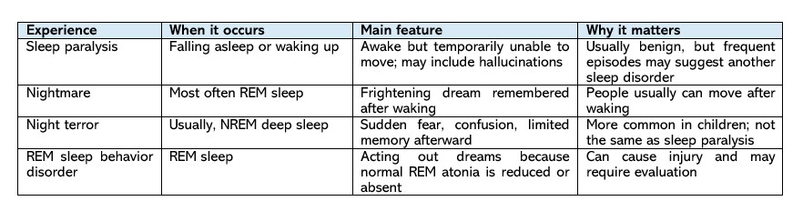
Quick comparison: similar sleep experiences
About the Author:
Tharoon Balakrishnan is a senior neuroscience student in the School of Behavioral and Brain Sciences at The University of Texas at Dallas, Richardson, Texas. He authored this article as part of Professor Faisal R. Jahangiri's Sleep and Sleep Disorders course. Tharoon is passionate about neuroscience and is committed to translating complex scientific concepts into engaging, accessible content for a broad audience.
References
Cheyne, J. A., Rueffer, S. D., & Newby-Clark, I. R. (1999). Hypnagogic and hypnopompic hallucinations during sleep paralysis: Neurological and cultural construction of the night-mare. Consciousness and Cognition, 8(3), 319–337. https://doi.org/10.1006/ccog.1999.0404
Cleveland Clinic. (2024, July 23). Multiple Sleep Latency Test (MSLT). https://my.clevelandclinic.org/health/diagnostics/multiple-sleep-latency-test-mslt
Denis, D., French, C. C., & Gregory, A. M. (2018). A systematic review of variables associated with sleep paralysis. Sleep Medicine Reviews, 38, 141–157. https://doi.org/10.1016/j.smrv.2017.05.005
Farooq, M., & Anjum, F. (2023). Sleep paralysis. In StatPearls. StatPearls Publishing. https://www.ncbi.nlm.nih.gov/books/NBK562322/
Harvard Medical School Division of Sleep Medicine. (n.d.). Science of sleep: What is sleep? Retrieved July 20, 2026, from https://sleep.hms.harvard.edu/education-training/public-education/sleep-and-health-education-program/sleep-health-education-47
Jahangiri, F. R. (2026a). Sleep cycle: Physiology in sleep [Lecture slides]. NSC 4320, University of Texas at Dallas.
Jahangiri, F. R. (2026b). Electromyography (EMG) [Lecture slides]. NSC 4320, University of Texas at Dallas.
Jahangiri, F. R. (2026c). Sleep disorders [Lecture slides]. NSC 4320, University of Texas at Dallas.
Jahangiri, F. R. (2026d). Electroencephalography (EEG) [Lecture slides]. NSC 4320, University of Texas at Dallas.
Jahangiri, F. R. (2026e). Polysomnography [Lecture slides]. NSC 4320, University of Texas at Dallas.
Jalal, B. (2016). How to make the ghosts in my bedroom disappear? Focused-attention meditation combined with muscle relaxation (MR therapy): A direct treatment intervention for sleep paralysis. Frontiers in Psychology, 7, 28. https://doi.org/10.3389/fpsyg.2016.00028
Krahn, L. E., Arand, D. L., Avidan, A. Y., Davila, D. G., DeBassio, W. A., Ruoff, C. M., & Harrod, C. G. (2021). Recommended protocols for the Multiple Sleep Latency Test and Maintenance of Wakefulness Test in adults: Guidance from the American Academy of Sleep Medicine. Journal of Clinical Sleep Medicine, 17(12), 2489–2498. https://doi.org/10.5664/jcsm.9620
Sharpless, B. A., & Barber, J. P. (2011). Lifetime prevalence rates of sleep paralysis: A systematic review. Sleep Medicine Reviews, 15(5), 311–315. https://doi.org/10.1016/j.smrv.2011.01.007